
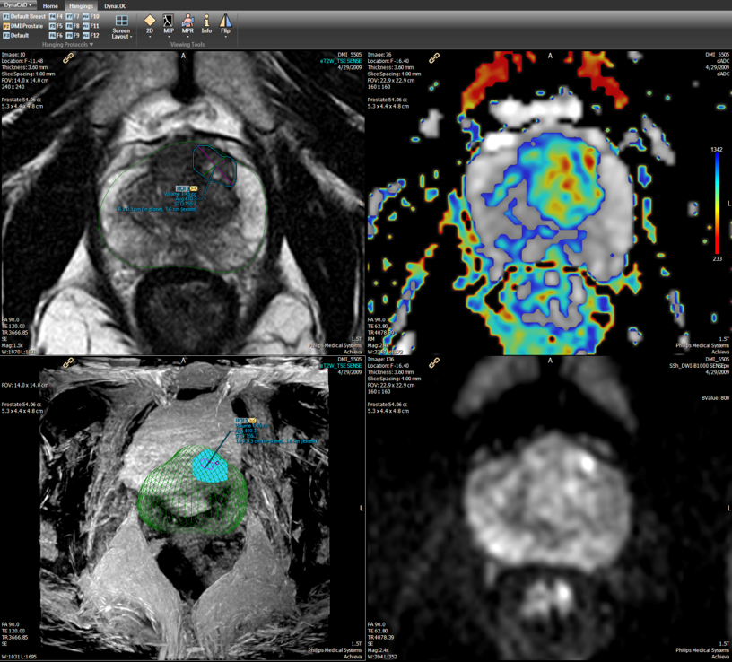
Multiparametric MRI
Multiparametric magnetic resonance imaging (MRI) refers to a sequence of imaging procedures that follow a specific set of protocols to visualize the prostate gland. Multiparametric MRI combines traditional anatomic imaging techniques with functional techniques to reveal detailed information about potential prostate cancers.
Anatomic Imaging
A basic, commonly-performed MRI sequence, T2-weighted imaging produces visualizations of the prostate gland and internal structures in high detail. Anatomic imaging can detect cancer within the prostate and assess spread of cancer beyond the prostate. Functional imaging exams are performed to more accurately identify the location of and gather other information about tumors within the prostate gland.
Diffusion-Weighted Imaging
Diffusion-weighted imaging (DWI) examines the movement of water molecules within the prostate to establish apparent diffusion coefficient (ADC) values. Because water molecules move differently through healthy tissue than they do through cancerous tissue, DWI can help physicians identify the boundaries of a tumor within the prostate gland. ADC values, which are produced automatically using specialized software, can also help physicians to assess presence, extent and aggressiveness of a tumor.
Dynamic Contrast-Enhanced
In dynamic contrast-enhanced (DCE) imaging, a contrast agent is administered prior to an MRI exam. MRI is then used to monitor concentrations of the contrast agent as it travels through blood vessels to prostate tissue. Because cancerous tissue and healthy tissue absorb the contrast agent at different rates, DCE imaging reveals information about a tumor’s location and extent of a tumor.
Benefits of Multiparametric MRI
Compared with conventional diagnostic tools used in prostate cancer detection and diagnosing, multiparametric MRI is proving to be useful in multiple phases of a more reliable option when it comes to determining which men would benefit from biopsy. Paired with prostate-specific antigen (PSA) screening, multiparametric MRI can be used to evaluate elevated and rising PSA levels prior to biopsy. Additionally, multiparametric MRI images can be used to guide biopsy needles with greater accuracy than other imaging techniques and can provide information to assist prostate cancer staging and treatment planning.
Preparing for a Prostate MRI Exam
- Drugs & Medications – Patients should be sure to inform RadNet staff what medications they are taking. Unless otherwise instructed by their physicians, patients should follow normal routines for taking prescribed drugs and medications.
- Food & Drink – Patients will be asked to refrain from eating and/or drinking coffee in the 4 hours prior to a prostate MRI in order to provide optimal image quality. Patients should also use the bathroom, emptying both their bladders and bowels, shortly before the exam begins.
- Clothing – Patients may be asked to change into a medical gown for their exam. For this reason, we recommend patients where comfortable, loose-fitting clothing. Patients will also be asked to remove any metallic jewelry and accessories, including dentures, eyeglasses and hearing aids.
- Sexual Activity – Patients should refrain from ejaculating in the 48 to 72 hours prior to a prostate MRI to ensure best possible visualization of the seminal vesicles.
The Day of a Prostate MRI Exam
- Arrival – Patients should arrive at the imaging facility at least 15 minutes before the scheduled start of their prostate MRI exam. This provides time to fill out any necessary paperwork, to change into appropriate exam clothing and to exchange questions and answers with one of our radiologic technicians.
- Scanning – The exam itself will take place in a private room equipped with a 3T MRI machine. The MRI machine is a large, cylindrical tube with an opening in the center. Patients will be asked to lie on their back on an exam table that slides in and out of the MRI machine. A radiologic technician helps to position patients correctly and to ensure that they are comfortable. The radiologic technician will then leave the room, but will be available to talk with patients via intercom. During the exam, the table on which you are positioned slides into the MRI machine. The machine produces a loud clicking noise during the procedure, but causes no pain. Patients can expect their prostate MRI exams to take around 45 minutes.
- Contrast Agent – A contrast agent introduced via a small IV catheter in a vein will be used for a portion of the exam. This helps to highlight internal organs and provides valuable information to radiologists. A blood test may be administered prior to a prostate MRI exam to ensure it will be safe to use.
Following a Prostate MRI Exam
- Recovery – Patients can generally resume normal activities as soon as their exam is complete. Physicians or radiologic technicians may provide instructions for recovery to some patients.
- Results – A radiologist interprets prostate MRI exams and returns results to the patient’s referring physician. Based on these results, patients and their physicians can develop a course of action based on patient preferences and individual health.